Welcome page
Welcome to the MRI labs of the Institutes of Radiology and Neuroradiology!
In the MRI labs of the Institute of Radiology (Head: Prof. Michael Uder) and the Institute of Neuroradiology (Head: Prof. Arnd Dörfler) in Erlangen, we develop new imaging techniques for magnetic resonance imaging . We test these methods intensively and in close cooperation with physicians.
Projects
Again and again, we offer exciting Bachelor, Master and PhD projects.
A particularly nice feature of research in MRI is the wide range of possible topics:
- Basic aspects of MR physics
- Theoretical considerations
- Computer simulations
- Implementation of new MR sequences
- At clinical MR-scanners (0.55 T to 7 T)
- At preclinical MR-scanners (7 T)
- Validations in measurement phantoms and healthy subjects
- Translation into clinical studies together with clinical partners and the focus areas:
- Oncology
- Immunology
- Renal and circulation research
- Dementia and neuroscience
- Muscular diseases
- Medical technology
- Advanced evaluation methods
- Image reconstruction
- Classic image processing
- Neural networks / AI
Our MRI labs have different thematic focuses (see Lab pages). We work closely together on many projects and benefit from our different strengths and expertises.
Contact us if you are interested!
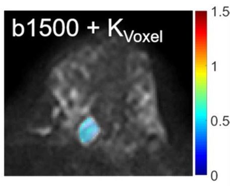 New MR techniques for breast cancer imaging (developed by our PhD student Mona Pistel) |
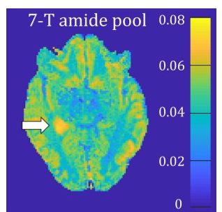 New MR methods for contrast agent free imaging of epilepsy-related brain tumors (developed by our PhD students Angelika Mennecke and Moritz Fabian) |
Teaching
We offer a wide range of courses. They prepare you optimally for projects in the field of MRI research, but also offer the opportunity to acquire comprehensive knowledge in the field of MRI, which will be of great benefit to you in other research projects and in industry.
We look forward to welcome you to our courses!
 Lecture MRI 1 with Prof. Dr. Frederik Laun |
Ultra High Field Imaging
A lot helps a lot. Sometimes this is indeed the case. In MRI, the signal-to-noise ratio increases with magnetic field strength. While clinical tomographs are usually operated with field strengths of 1.5 T or 3 T, clinical devices of the latest generation are also available with 7 T. Unfortunately, a higher field strength is often accompanied by increased image artifacts. For example, the wavelength of the transmitting field decreases with higher field strength, so that the images are inhomogeneous in intensity. We are working on overcoming these technical difficulties, establishing various MR techniques to 7 T scanners and being able to apply them in acceptable measurement time.
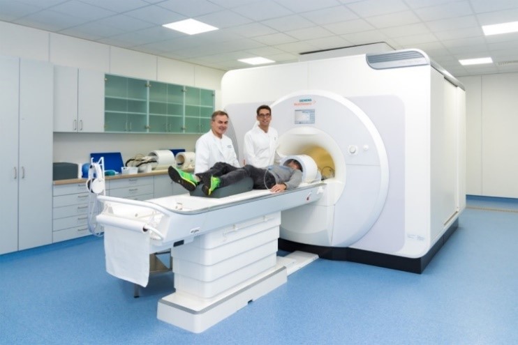
The 7 T Magnetom Terra Tomograph of the University Hospital Erlangen
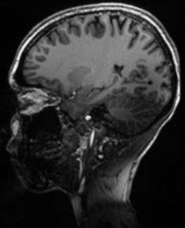 |
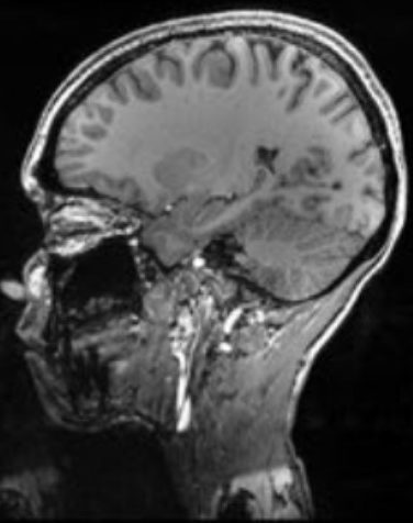 Top: Inhomogeneous appearance due to the short transmit field wavelength at 7 T. Bottom: Parallel transmit techniques (ptx) increase the homogeneity of the intensity. (developed by our former PhD student Jürgen Herrler) |